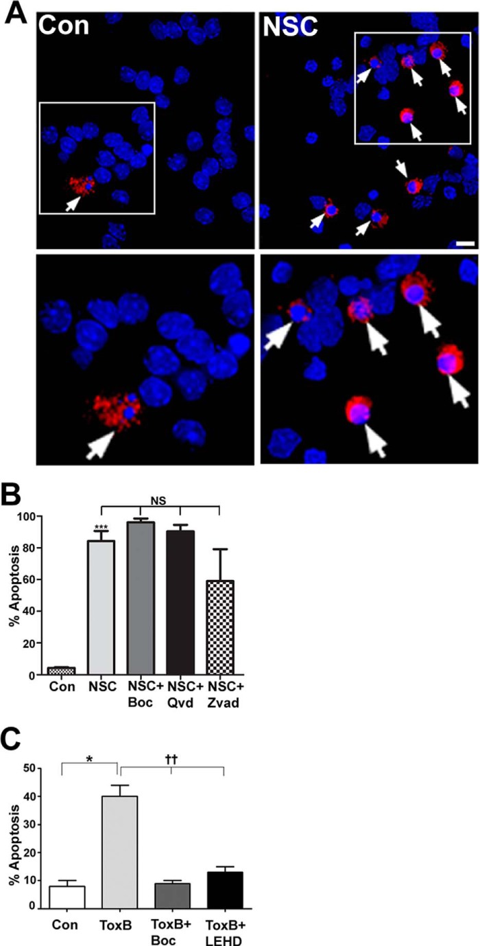
FIGURE 3.
CGN death induced by NSC23766 is largely caspase-independent, unlike Toxin B-induced apoptosis. A, cells were incubated for 48 h in either control medium alone (Con) or medium containing NSC23766 (NSC, 200 μm). Cells were then fixed, and their nuclei were stained with DAPI. Active caspase-3 was visualized using a polyclonal antibody and a Cy3-conjugated secondary antibody. NSC23766 treatment causes increased activation of caspase-3 in CGNs (arrows). Scale bar = 10 μm. B, cells were incubated for 48 h in either control medium or medium containing NSC23766 (200 μm) with or without BOC (50 μm), QVD (20 μm), or ZVAD (10 μm). The percentage of cell viability was measured by nuclear morphology. Data represent the mean ± S.E. of three independently performed experiments. ***, p < 0.001; NS, no significant difference between NSC and NSC cotreated with caspase inhibitors individually (BOC, QVD, or ZVAD). C, cells were incubated for 24 h in either control medium or medium containing ToxB (60 ng/ml) with or without BOC (50 μm) or LEHD (100 μm, a caspase-9 inhibitor). Data represent the mean ± S.E. of four independently performed experiments. *, significantly different from control (p < 0.05); ††, significantly different from ToxB (p < 0.01).

