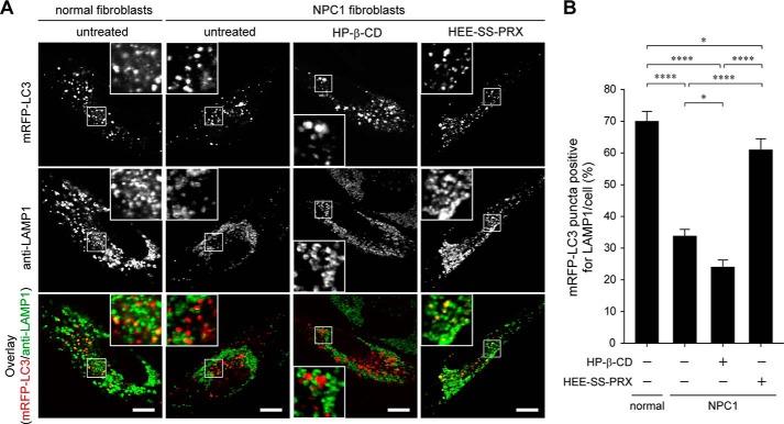
FIGURE 7.
Biocleavable polyrotaxanes facilitated the colocalization of LC3 and LAMP1. A, CLSM images of mRFP-LC3 (first row) and anti-LAMP1 staining (second row) and overlay images (third row) in normal and NPC1 fibroblasts expressing mRFP-LC3 treated with HP-β-CD (10 mm) and HEE-SS-PRX (1 mm β-CD) for 24 h (scale bars, 20 μm). B, colocalization percentage of mRFP-LC3-positive puncta to anti-LAMP1-positive puncta. The values are expressed as the mean ± S.E. (error bars) of 30 cells (*, p < 0.05; ****, p < 0.001).

