FIGURE 8.
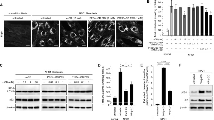
The effect of α-CD-threaded PRXs and other β-CD derivatives on autophagic flux. A, filipin staining for cholesterol in normal and NPC1 fibroblasts treated with α-CD (10 mm), PEG/α-CD PRX (1 mm α-CD), and P123/α-CD PRX (1 mm α-CD) for 24 h (scale bars, 20 μm). B, the amount of total cholesterol in normal and NPC1 fibroblasts treated with α-CD, PEG/α-CD PRX, and P123/α-CD PRX at various concentrations for 24 h (n = 3) (*, p < 0.05 against untreated NPC1 fibroblasts; NS, not significant). C, immunoblot analysis for LC3, p62, and β-actin in the NPC1 fibroblasts treated with α-CD, PEG/α-CD PRX, and P123/α-CD PRX at various concentrations for 24 h. D, the amount of total cholesterol in the normal and NPC1 fibroblasts treated with DM-β-CD (1 mm) and HP-β-CD (1 mm) for 24 h (n = 3) (*, p < 0.05; **, p < 0.01). E, the amount of cholesterol extracted from the plasma membrane to the culture medium after the treatment with DM-β-CD (10 mm) and HP-β-CD (10 mm) for 2 h at 4 °C (n = 3) (****, p < 0.001). F, immunoblot analysis for LC3, p62, and β-actin in the NPC1 fibroblasts treated with DM-β-CD (1 mm) and HP-β-CD (1 mm) for 24 h. The values are expressed as the mean ± S.D. (error bars).

