FIGURE 2.
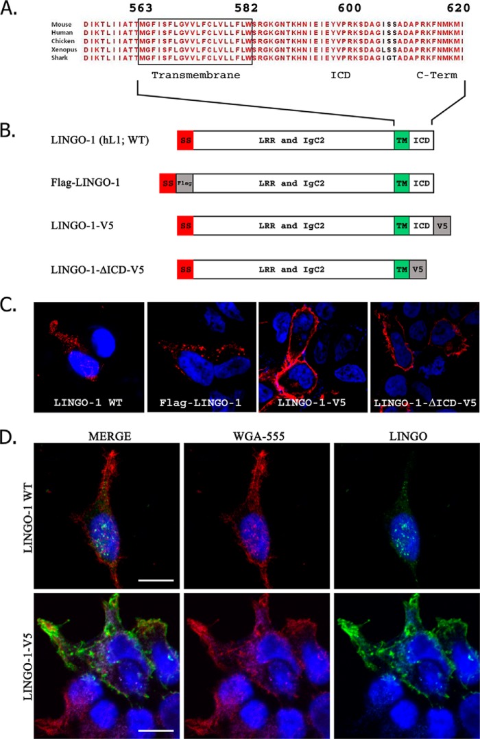
The LINGO-1 C terminus is essential for proper subcellular localization. A, illustration of the conservation of the LINGO-1 C terminus across vertebrate evolution. B, structure of various LINGO-1 constructs employed. h, human; SS, signal sequence; LRR, leucine-rich repeat; TM, transmembrane domain. C, subcellular distribution of wild-type (WT) and mutant LINGO-1 expressed in transiently transfected HEK293 cells imaged by confocal immunofluorescence microscopy. LINGO-1 detected by anti-LINGO, anti-FLAG, or anti-V5 antibody is shown in red. DAPI nuclear counterstain is shown in blue. Wild-type LINGO-1 or LINGO-1 bearing an N-terminal FLAG tag was expressed as apparent intracellular puncta, whereas LINGO-1 with an ICD deletion or bearing a C-terminal V5 tag trafficked to the cellular membrane. D, confocal immunofluorescence microscopy comparing distribution of LINGO-1 with distribution of lectin-labeled plasma membrane. HEK293 cells transiently transfected with LINGO-1 or LINGO-1 bearing a C-terminal V5 tag were briefly fixed and then reacted with fluorescently tagged Alexa Fluor 555-conjugated WGA (red), after which membranes were permeabilized by exposure to Triton X-100-containing buffer, and cells were immunostained with anti-LINGO-1 antibody (green). DAPI nuclear stain is shown in blue. LINGO-1-V5 co-localized extensively with Alexa Fluor 555-conjugated WGA-labeled plasma membrane, whereas LINGO-1 exhibited very little co-localization with Alexa Fluor 555-conjugated WGA. Scale bars = 10 μm.

