FIGURE 5.

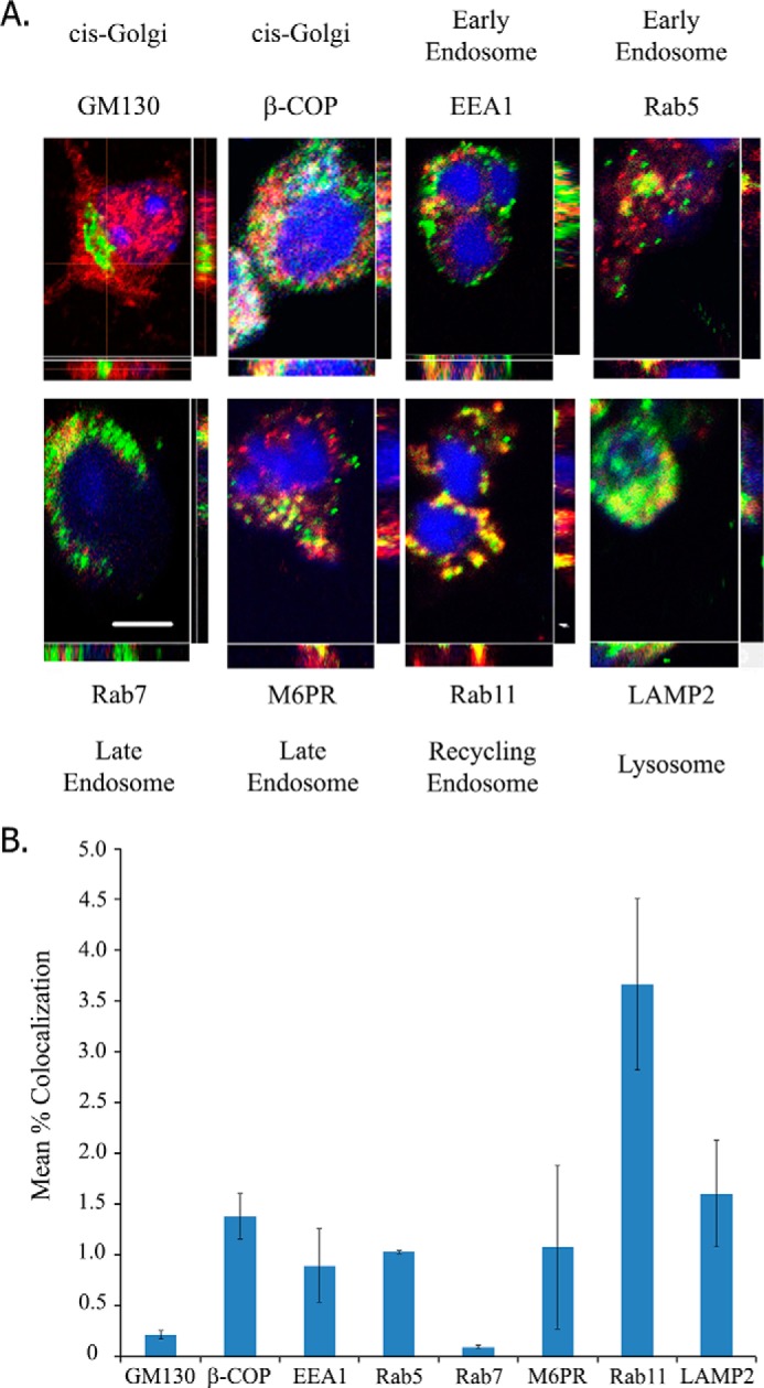
Neuronal LINGO-1 localizes to subcellular membrane compartments of secretory and endocytic pathways. A, confocal images of immunofluorescently stained LINGO-1 (red) and various membrane compartment markers (green) in cultured mouse cortical neurons. Images are maximum intensity projections with variation of density across orthogonal confocal image slices also depicted. Nuclei were stained with DAPI (blue). Membrane compartment markers include EEA1 (early endosomes), Rab5 (early endosomes), GM130 (cis-Golgi), β-COP (vesicles involved in anterograde and retrograde transport to/from the Golgi), mannose 6-phosphate receptor (M6PR; late endosomes), Rab7 (late endosomes), Rab11 (recycling endosomes), and LAMP2 (lysosomes). Scale bar = 10 μm. B, quantitation of LINGO co-localization with markers of various membrane compartments, assessed as percent of pixels displaying immunofluorescence signals for LINGO and each marker. Of the markers imaged, the greatest LINGO co-localization was observed with Rab11. Values are expressed as mean percent co-localization ± S.E. Rab11, n = 197 cells; GM130, n = 52 cells; β-COP, n = 96 cells; EEA1, n = 266 cells; Rab5, n = 343 cells; Rab7, n = 39 cells; mannose 6-phosphate receptor, n = 138 cells; LAMP2, n = 106 cells.
