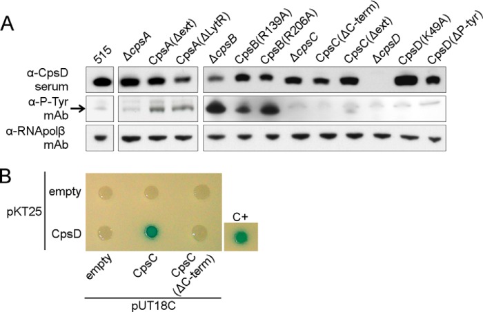
FIGURE 2.
CpsD and CpsB form an autokinase/phosphatase pair. A, Western blots showing CpsD (α-CpsD), tyrosine phosphorylation of CpsD (α-P-Tyr mAb), and the loading control RNA polymerase subunit β (α-RNApolβ mAb) in total protein extracts from 515 WT and the cps mutant strains. The band corresponding to the phosphorylated CpsD is indicated by an arrow. B, bacterial two-hybrid analysis of CpsC and CpsD. T25-CpsD was tested for interaction with T18-CpsC and T18-CpsC(ΔC-term). Control plasmids (T18 and T25) were tested together with fusion proteins as negative controls. The positive control used was the leucine zipper GCN4 fused to the T25 and T18 fragments (28). The formation of blue colonies indicates a protein-protein interaction.

