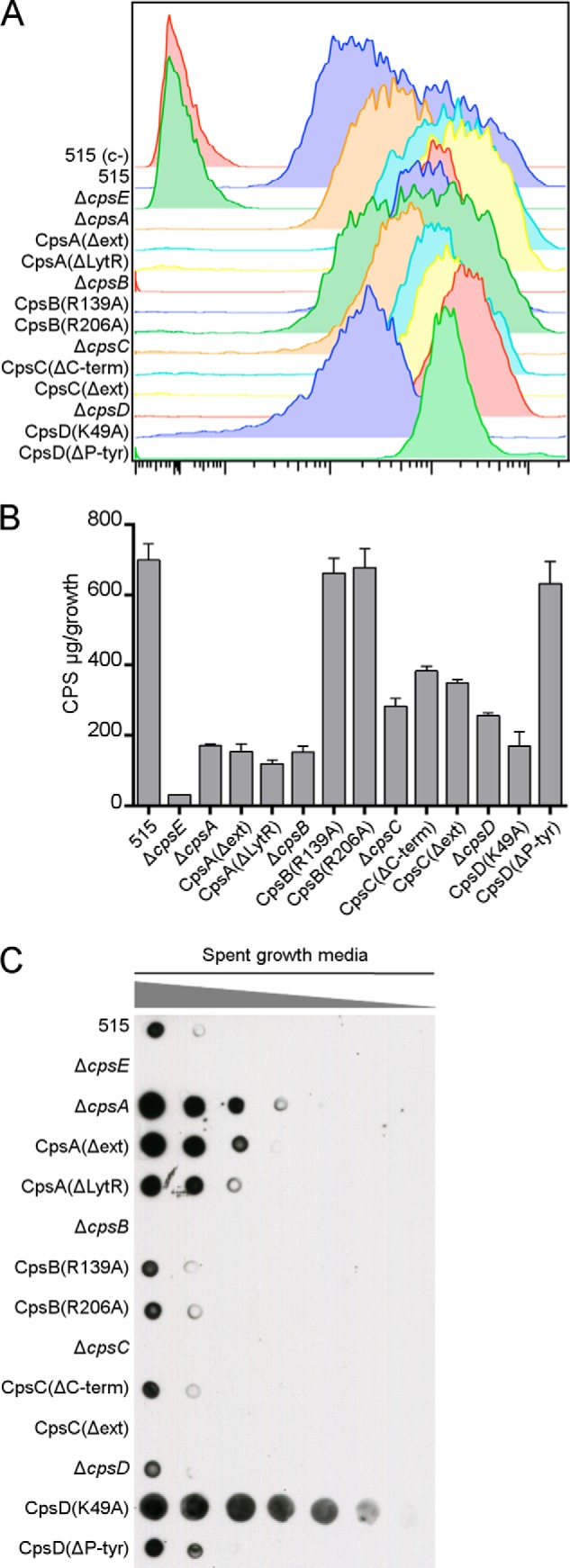
FIGURE 3.

Differences in CPS localization in mutant strains. A, WT and cps mutant strains were incubated with a primary α-CPSIa mAb followed by a secondary goat α-mouse antibody conjugated to allophycocyanin and analyzed by flow cytometry. The unencapsulated ΔcpsE strain was included as a negative control. B, CPS from bacterial pellets was quantified by a resorcinol assay. Bars, means of three independent experiments performed with triplicate samples. Error bars, S.D. C, dot blot showing serial dilutions (1:2) of spent growth media spotted on a nitrocellulose membrane and probed with an α-CPSIa mAb.
