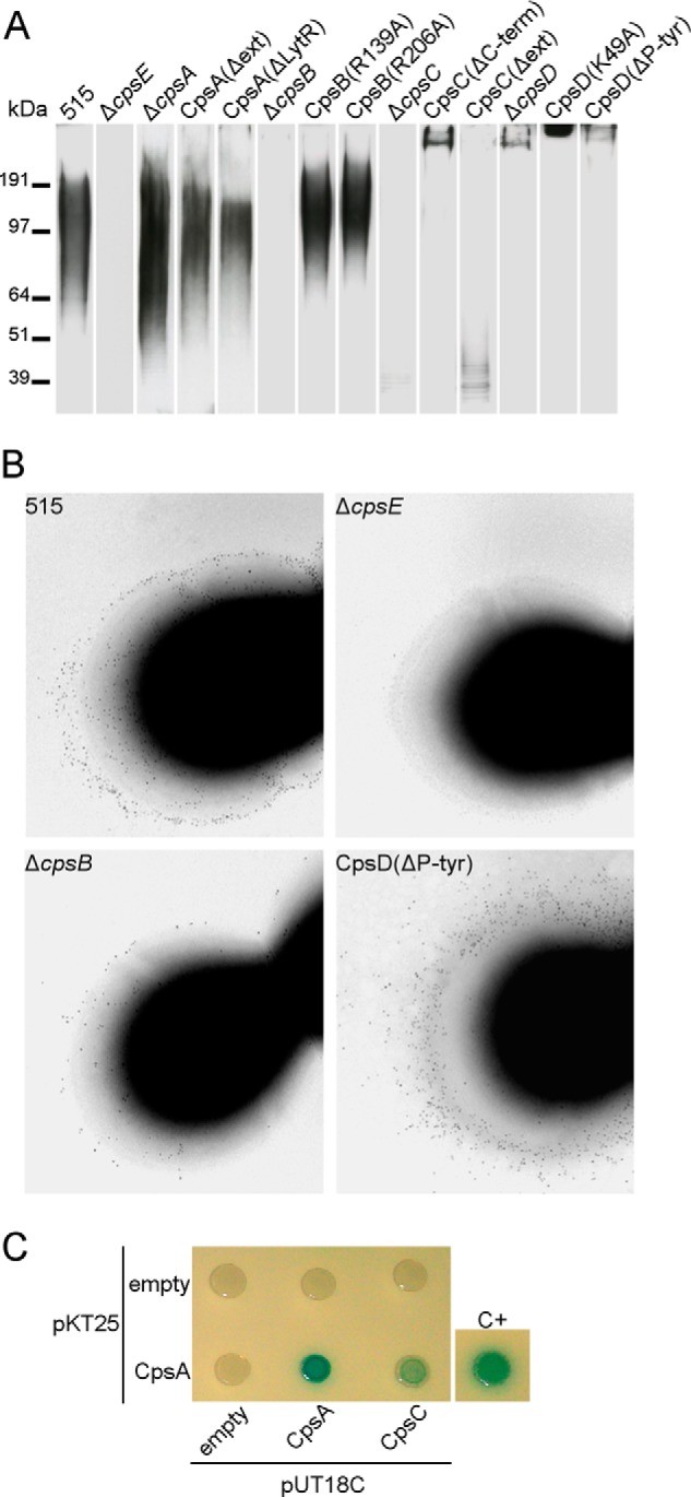
FIGURE 4.

CPS length differences in the WT and cps mutant strains. A, Western blot of CPS bacterial surface extracts (top). CPS was detected with an α-CPSIa mAb. A protein molecular weight marker is included for approximate comparison. Lanes were exposed for different times, in order to permit visualization of CPS from all strains. B, immunogold transmission electron microscopy on whole bacteria using an α-CPSIa mAb as primary antibody and a secondary gold bead-conjugated antibody. Bacterial strains are indicated. C, bacterial two-hybrid analysis. T25-CpsA was tested for interaction with T18-CpsA and T18-CpsC. T18 plasmids were used as negative control. The positive control (C+) is the GCN4 leucine zipper protein provided by the manufacturer in plasmids pKT25-zip and pUT18C-zip (28). Formation of blue colonies indicates that a protein-protein interaction occurred.
