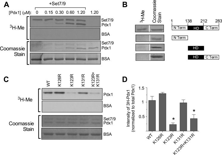
FIGURE 1.
Pdx1 methylation by Set7/9 in vitro. A, a methylation assay in vitro using recombinant Set7/9, [3H]AdoMet and increasing concentrations of Pdx1 protein was performed, and then reactions were subjected to polyacrylamide gel electrophoresis. The first and second panel show fluorography for 3H, and the third and fourth panels show corresponding Coomassie staining of the same gel. B, methylation assay in vitro using recombinant Set7/9, [3H]AdoMet, and full-length or truncated Pdx1 proteins was performed, followed by polyacrylamide gel electrophoresis. A schematic of truncated mutants of Pdx1 is shown in the right panel, and corresponding 3H fluorography and Coomassie stains are shown in the left panel. Term, terminus; HD, homeodomain. C, methylation assays in vitro using WT and mutated Pdx1 proteins were performed with corresponding quantitation of methylated Pdx1 protein intensities (normalized to total Pdx1 protein by Coomassie staining). All images shown are representative of at least three experiments. *, p < 0.05 compared with wild-type Pdx1.

