FIGURE 6.
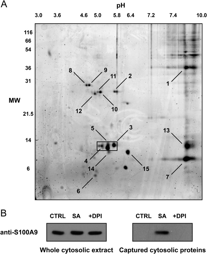
Identification of cytosolic biotin hydrazide-derivatized carbonyl proteins in phagocytic neutrophils by two-dimensional gel electrophoresis and MS. Cytosolic extracts of neutrophils treated with S. aureus at a ratio of 20:1 were derivatized with biotin hydrazide, and the biotinylated proteins were captured with magnetic streptavidin-coated beads. A, biotin hydrazide-derivatized carbonyl proteins captured from the beads were separated by two-dimensional electrophoresis with 12% SDS-PAGE in the second dimension. Gels were then stained for total protein with SYPRO Ruby. The gel is representative of seven experiments, and the boxed region highlights the 14-kDa proteins that were predominantly captured over the seven experiments. MW, molecular weight. B, the whole cytosolic extract before bead capture (left panel) and the biotinylated proteins captured with magnetic streptavidin-coated beads (right panel) were separated by electrophoresis and Western blotted with a specific MRP-14 antibody. The blots are representative of three experiments. SA, S. aureus (20:1).

