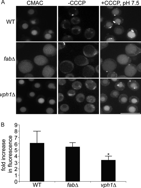
FIGURE 3.

Vacuole-targeted pHluorin suggests that fab1Δ vacuoles are acidic. A, wild-type (WT), fab1Δ, and vph1Δ yeast cells carrying a genomic copy of Mup1-pHluorin were grown in the presence of methionine and stained with CMAC to label vacuoles. Cells were then mounted on concanavalin-coated slides and imaged live by spinning disc confocal microscopy before and after the addition of CCCP, a proton ionophore, in media set to pH 7.5. Scale bar = 10 μm. B, to measure changes in pHluorin intensity, images were obtained using epifluorescence ratiometric imaging. Regions of interest were then drawn over vacuoles to obtain the fluorescence intensity of pHluorin before and after CCCP addition. Fluorescence intensities were then background-corrected, and the fluorescence ratio after and before CCCP addition was calculated. Shown is the mean ± S.E. for wild-type (five experiments with a total of 514 vacuoles), fab1Δ (five experiments with a total of 372 vacuoles), and vph1Δ (three experiments with a total of 324 vacuoles) cells. Using ANOVA and Tukey's post hoc test, we show that there was no statistical difference between wild-type and fab1Δ cells, but there was a significant difference (*) between vph1Δ cells against control or fab1Δ cells (p < 0.01 for wild-type and p < 0.05 for fab1Δ).
