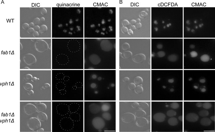
FIGURE 4.
Vacuolar accumulation of quinacrine and cDCFDA. The vacuoles of wild-type, fab1Δ, vph1Δ, and fab1Δ vph1Δ double mutant yeast cells were labeled with CMAC and with either 200 μm quinacrine for 10 min (A) or 50 μm cDCFDA for 1 h (B). A, dashed lines outline cells with little to no quinacrine signal. Corresponding differential interference contrast (DIC) images are also shown. Live cell imaging was done with an epifluorescence microscope (scale bar = 10 μm).

