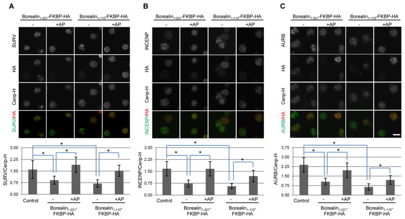
Figure 2. Borealin dimerization regulates CPC abundance at centromeres.
HeLa M cells were transfected with the indicated constructs and then exposed to thymidine for 24 hours to synchronize in S phase. At the time of thymidine removal, taxol was added in combination with AP20187 (+AP) or DMSO (−). Cells were fixed 16 hours later and analyzed by immunofluorescence. Intensity of centromere staining of Survivin (A), INCENP (B) and Aurora B (C) was quantified relative to the kinetochore marker CenpH (scale bar = 20μm). Bars represent standard deviation from averages of at least 50 centromere signals from 10 different cells per condition. *p<0.05 using a student’s t-test.

