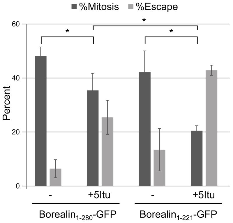
Figure 9. Checkpoint function of a kinetochore-proximal CPC pool.
HeLa M cells were transfected and synchronized into S-phase by 24-hour treatment with 2mM thymidine. Following synchronization, cells were released into taxol for 16 hours and time-lapse imaging was performed immediately after adding either DMSO (−) or 5Itu. The percentage of Borealin-GFP expressing cells remaining in mitosis or having escaped mitosis by 15-hours after 5Itu treatment is shown. Bars represent standard error from three independent transfections. *p<0.05 from a student’s t-test.

