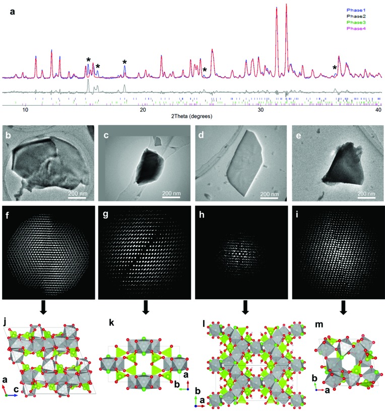
Figure 2.
(a) Rietveld refinement against the PXRD pattern using the four phases of a multiphase Ni–Se–O–Cl sample determined from RED data (λ = 1.5418 Å). Unindexed peaks are marked by asterisks (*). TEM images showing crystals of (b) Phase 1, (c) Phase 2, (d) Phase 3 and (e) Phase 4 used for RED data collection. The corresponding three-dimensional reciprocal lattices of (f) Phase 1, (g) Phase 2, (h) Phase 3 and (i) Phase 4 reconstructed from RED data. Structure models of (j) Phase 1 (NiSeO3), (k) Phase 2 (Ni3Se4O10Cl2), (l) Phase 3 (Ni5Se6O16Cl4H2) and (m) Phase 4 (Ni5Se4O12Cl2) determined from RED data. The NiO4Cl2, NiO5Cl and NiO6 octahedra are in grey, SeO3 trigonal pyramids and Se atoms in yellow green, O atoms in red, Cl atoms in green and H atoms in brown. More information is given by Yun et al. (2014 ▶), from where this image is reproduced.

