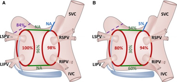
Figure 3.

Success rate of surgical thoracoscopic epicardial radiofrequency isolation of pulmonary veins, linear ablation lines connecting both superior and inferior pulmonary veins, and the trigone line connecting the right superior pulmonary vein across the left atrial roof toward the noncoronary aortic cusp as they were assessed (A) immediately after the ablation during surgery and (B) during the electrophysiological examination 6 to 8 weeks following the index procedure. Percentage of deployed left atrial appendage clips is also depicted (violet color). IVC indicates inferior vena cava; LIPV, left inferior pulmonary vein; LSPV, left superior pulmonary vein; NA, not applicable; RIPV, right inferior pulmonary vein; RSPV, right superior pulmonary vein; SVC, superior vena cava.
