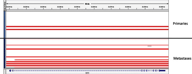
Figure 1.

E2F3amplification in primary tumors vs. metastases. Analysis of E2F3 gene copy number data using IGV with each row representing a single tumor sample. Primary tumor samples are arrayed above the black line and metastases below it. On the left side of the diagram, the light blue boxes represent primary tumor samples and the dark blue boxes represent metastases. Red bars represent amplification (log2 copy number ratio >0.8).
