
Figure 2.
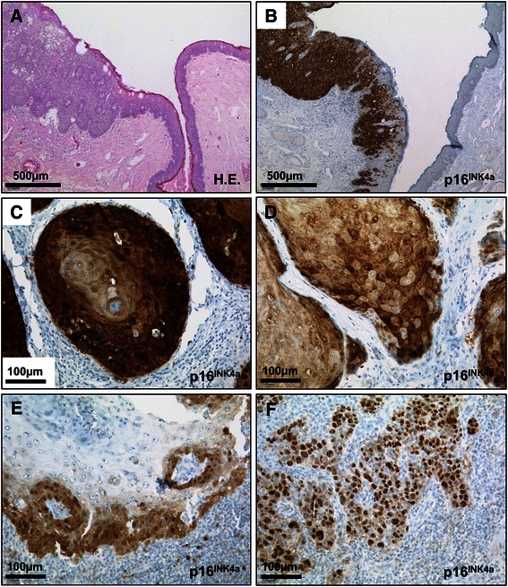
Representative microphotographs of pSCC specimens. A and B, penile squamous cell carcinoma in situ displaying strong, confluent expression of p16INK4a. Note ascending p16INK4a positivity in non-malignant epithelium adjacent to carcinoma. C-F, variety of p16INK4a staining patterns in pSCC: C, strong, confluent staining in invasive keratinizing carcinoma; D, diffuse and E, focally scattered positivity for p16INK4a; F, almost exclusively nuclear immunostaining. Scale bars as indicated; H.E., haematoxylin-eosin; HR-HPV, high-risk human papillomavirus; pSCC, penile squamous cell carcinoma.