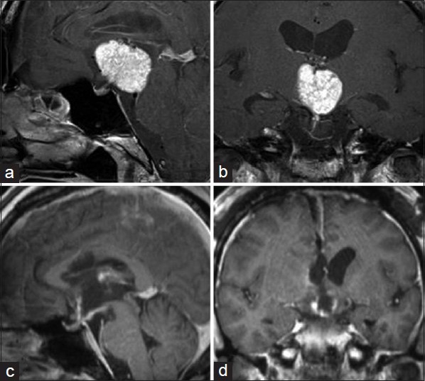
Figure 1.

Gadolinium (Gd)-enhanced magnetic resonance (MR) images. In the preoperative images, the tumor was homogeneously enhanced by Gd, which extended to the dorsal part in the third ventricle (a and b). The MR images obtained 5 days after the operation. The tumor was totally removed with mild enhancement of the ventricular wall reflecting postoperative changes (c and d)
