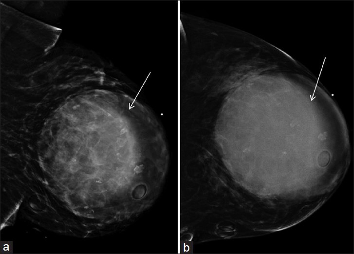
Figure 1.

46-year-old female with history of neurofibromatosis Type 1 presented with left breast mass diagnosed as metaplastic breast carcinoma. Mediolateral (a) oblique and (b) craniocaudal views of left breast mammogram demonstrate a large, relatively circumscribed mass (arrows) adjacent to a dot-shaped radiopaque skin marker to indicate the palpable area in the central left breast. It measured approximately 10 × 10 × 9 cm. Circular-shaped radiopaque skin markers were placed to indicate the skin lesions (neurofibromas) which are consistent with patient's known neurofibromatosis.
