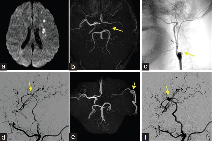
Figure 1.

(a) Diffusion-weighted MR image at onset indicating acute watershed infarction due to hemodynamic insufficiency. (b and c) MR angiogram (b) and lateral DSA of the left common carotid artery (c) at the onset showing occlusion of the left ICA at the cervical portion (arrow). (d) Lateral DSA of the left common carotid artery after the first anastomosis in which the blood flow from the STA perfuses both proximal and distal to the anastomotic site (arrow). (e and f) MR angiogram (e) and lateral DSA of the left common carotid artery (f) performed 2.5 years after the first anastomosis showing de novo aneurysm at the anastomotic site (arrow)
