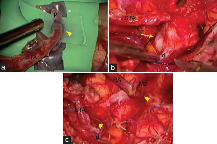
Figure 2.

(a) Intraoperative photograph of the first STA-MCA anastomosis (arrowhead). (b and c) Intraoperative photographs of the trapping of the aneurysm and second ECA-RA-M2 and STA-M4 anastomoses showing the de novo aneurysm (arrow) pretrapping view (b), and posttrapping and anastomoses (arrowhead) view (c). d: M4 portion of the MCA distal to the anastomosis; M2: M2 portion of the MCA; p: M4 portion of the MCA proximal to the anastomosis; RA: Radial artery; STA: Superficial temporal artery
