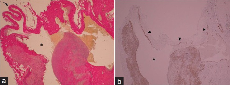
Figure 3.

Photomicrographs of resected de novo aneurysm. Left: Wall of the aneurysm dome (arrow) is thinned and consisted of collagen fibers. Asterisk = aneurysm lumen. Elastica van Gieson stain, original magnification ×20. Right: Fragmented smooth muscle cells (arrowhead) are detected throughout the circumference. Alpha-smooth muscle actin stain, original magnification ×20
