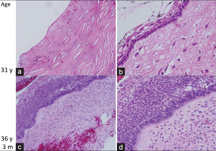
Figure 4.

Pathological findings. (a and b) Histopathology at 31 years of age. The tumor is covered with prickle cells, and a single layer of nonatypical basal cells and cholesterin crystals is present in the interstitial tissue. The diagnosis was adamantinomatous craniopharyngioma. (c and d) Histopathology at 36 years and 3 months of age. Densely packed squamous cells and stratification of basal cells with an atypical appearance are seen. The features of adamantinomatous type craniopharyngioma are no longer apparent. (c–d) Hematoxylin and eosin staining at the original magnification
