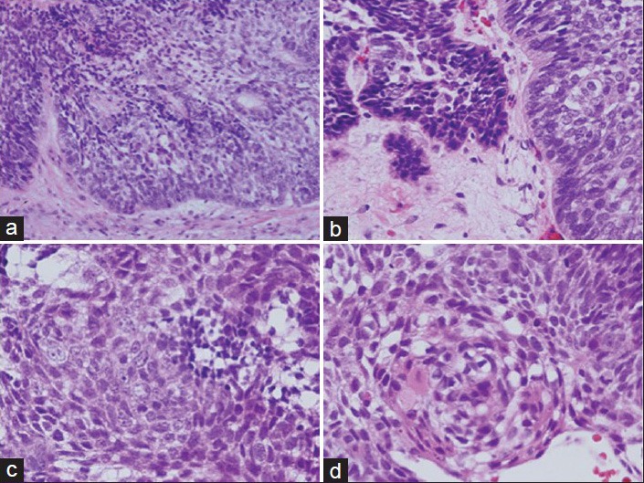
Figure 5.

Pathological findings of a specimen from the transcranial and transsphenoidal dual surgeries at 36 years and 9 months of age. (a) The number of squamous cells has increased. (b) The lamina propria has collapsed, and infiltration of atypical cells is seen. (c) Tumor cells have enlarged nuclei and clarification of the nucleolus. (d) Parakeratosis and intercellular bridges are present in the tissue. Hematoxylin and eosin staining at the original magnification
