
Fig. 3.
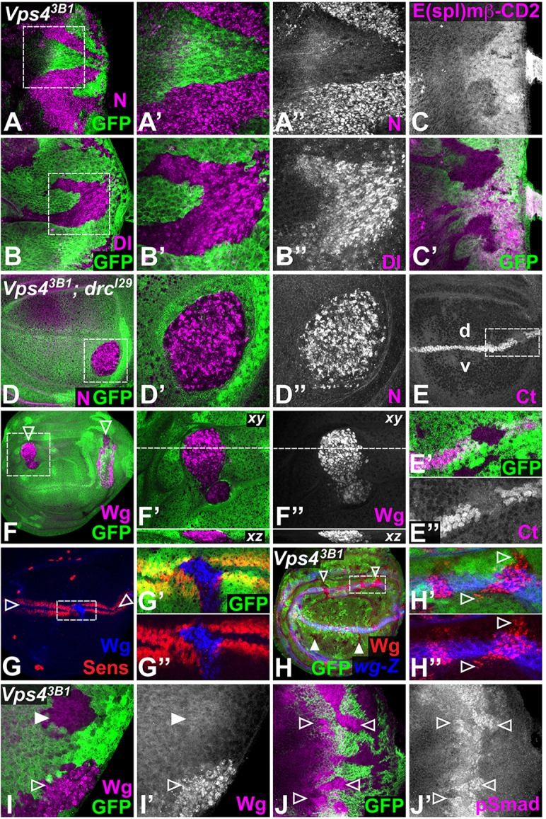
Loss of Vps4 affects multiple signaling pathways. Vps43B1 clones marked by the absence of GFP (green) in eye discs (A-C,I-J) or in DroncI29 (drcI29) homozygous wing discs (D-H). (A,B) Mutant cells accumulate high levels of N (A″, magenta in A,A′) and Dl (B″, magenta in B,B′) in large puncta (boxed regions are enlarged in A′,A″,B′,B″). (C) Expression of the Notch transcriptional reporter E(spl)-mβ-CD2 (C, magenta in C′) is decreased in Vps4 clones. (D) Mutant cells accumulate large N-positive puncta (D″, magenta in D,D′, the boxed region is enlarged in D′,D″). (E) The Notch transcriptional target Ct (E,E″, magenta in E′), normally expressed at the boundary between the dorsal (d) and ventral (v) compartments of the wing pouch, is lost in Vps4 clones (the boxed region is enlarged in E′,E″). (F) Wg (F″, magenta in F,F′) also accumulates in large puncta that span the depth of the cell in Vps4 clones (arrowheads; the boxed region is enlarged in F′,F″; xz sections are shown below). (G) The Wg target Sens (red) is expressed in two stripes (arrowheads) along the D-V boundary of the wing pouch, but is lost from Vps4 clones, despite accumulation of Wg (blue). The boxed region is enlarged in G′,G″. (H) Ectopic Wg (red) is observed in Vps4 clones in the vicinity of Wg-producing cells (open arrowheads), marked by wg-lacZ (blue), but not in more distant clones (filled arrowheads). The boxed region is enlarged in H′,H″. (I) In eye discs, Wg puncta (I′, magenta in I) are observed in clones along the lateral margins (open arrowhead) but not in more medial clones (filled arrowhead). (J) Vps4 clones display elevated levels of pSmad (J′, magenta in J) in the region of the morphogenetic furrow (arrowheads).