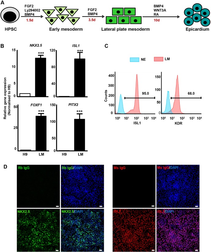
Fig. 1.
Induction of lateral plate mesoderm (LM). (A) Schematic of HPSC differentiation to LM and epicardium. HPSCs were differentiated to early mesoderm using FGF2, Ly294002 and BMP4 for 36 h, and subsequently to LM with FGF2 and BMP4 for 3.5 days. (B) Analysis of LM/splanchnic mesoderm markers in LM by qRT-PCR. Student's t-test, ***P<0.001. (C) Percentage of ISL1+ and KDR+ cells in LM determined by flow cytometry. H9-derived neuroectoderm (NE) was used as a negative control. (D) The majority of LM cells immunostained positive for NKX2.5 and ISL1. Rabbit and mouse IgG isotypes were used as negative controls. Scale bars: 100 μm.

