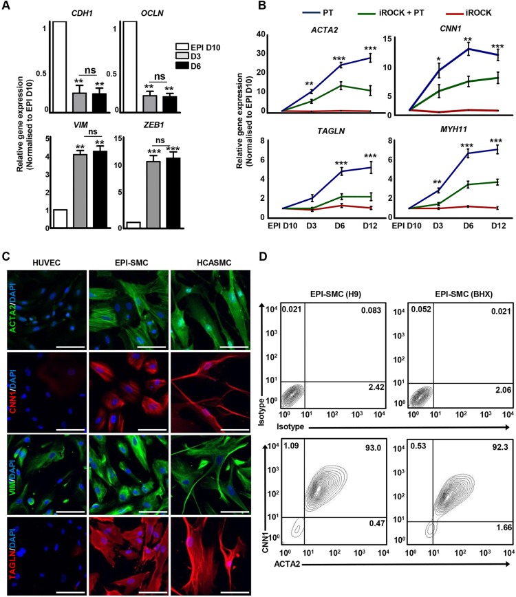
Fig. 5.
Epicardium-derived SMC differentiation in vitro. (A) Epithelial and mesenchymal marker expression in day 10 epicardial cells (EPI D10) and epicardium-derived SMCs (EPI-SMCs) after 3 (D3) and 6 (D6) days of differentiation with PDGF-BB and TGF-β1 (PT). ***P<0.001, **P<0.01. (B) SMC marker expression by qRT-PCR in EPI-SMCs differentiated with PT in the presence and absence of p160 Rho-kinase inhibitor (iROCK) after 3, 6 and 12 days of differentiation. Significant differences between PT and iROCK+PT are indicated in black. ***P<0.001, **P<0.01, *P<0.05. (C) EPI-SMCs after 12 days of PT treatment expressed mesenchymal (VIM) and SMC (ACTA2, CNN1 and TAGLN) markers, similar to human coronary artery SMCs (HCASMCs). SMC marker expression was absent in HUVECs. Scale bars: 100 μm. (D) Percentage of ACTA2+ and CNN1+ cells in H9 and BHX-derived EPI-SMCs. Mouse IgG isotypes served as negative controls.

