
Figure 1.
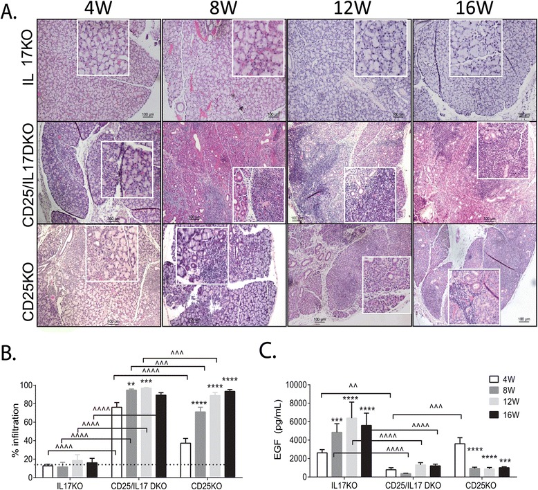
Accelerated and more severe dacryoadenitis in CD25/IL-17 DKO LG. (A) Representative images of H&E-stained LG from mice aged 4 to 16 weeks (W). The square is a higher magnification of the area underneath. Scale bar = 100 μm. (B) Percent area of lymphocytic infiltration measured in digital images of H&E-stained LG sections from IL-17KO, CD25/IL-17 DKO and CD25KO mice at 4, 8, 12, and 16 W. (C) EGF concentration measured in tear washings from different strains. * P <0.05;** P <0.01, *** P <0.001; **** P <0.0001 within strain comparison vs. 4 W. ^ P <0.05; ^^ P <0.01, ^^^ P <0.001; ^^^^ P <0.0001 interstrain comparison. CD25/IFN-γ DKO, CD25−/−IFN-γ−/−; CD25KO, CD25 knockout; DKO, double knockout; EGF, epidermal growth factor; H&E, hematoxylin and eosin; IL, interleukin; IL-17KO, IL-17 knockout; KO, knockout; LG, lacrimal gland.