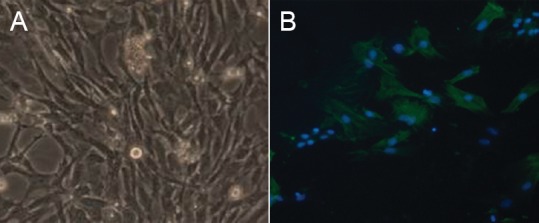
Figure 1.

Morphological identification of cultured Schwann cells (× 40).
(A) Phase contrast microscopy showed that Schwann cells were tightly arranged, spindle-shaped, narrow, with small nuclei. (B) Immuno-histochemical staining shows the myelin basic protein, expressed in Schwann cell bodies and processes, as green fluorescence (fluorescein isothiocyanate, FITC). Nuclei exhibited blue fluorescence (4′,6-diamidino-2-phenylindole, DAPI). The cytoplasm of fibroblasts was not stained, but their nuclei were stained blue.
