Figure 1.

Schematic diagram of the laminectomy site.
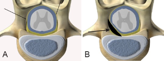
The first lumbar vertebra (A, indicated by black lines) and the self-made silica gel pad compressing the thirteenth thoracic ventral horn (B, arrow).

Schematic diagram of the laminectomy site.
The first lumbar vertebra (A, indicated by black lines) and the self-made silica gel pad compressing the thirteenth thoracic ventral horn (B, arrow).