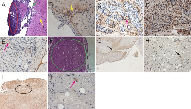
Figure 6.
Histological changes to xenografts after transplantation of CD133+ astrocytoma cells.
(A) At 8 weeks after cell transplantation, xenografts were observed outside the cerebral cortex of CD133+ mice by hematoxylin-eosin staining. A large tumor nodule protrudes from the surface of cortex, in which there are atypical cells of different sizes (red oval circle). There are several vessels that supply blood to the tumor nodule in the juncture between the nodule and cortex (as shown by green arrows). Some diffuse astrocytoma cells like pseudopodia infiltrated the surrounding subcortical white matter (as shown by yellow arrows). Original magnification: × 100. (B) Many glioma cells on the surface of cortex of CD133+ mouse showed strong GFAP-positive expression, several cells infiltrated deeply into the white matter (pointed by arrow). Original magnification: × 400. (C) Nestin immunoreactivity was seen in vascular cavity, wall, and extracellular matrix (arrow). Original magnification: × 200. (D) Strong Notch immunoreactivity was seen in the cell cytoplasm. Proliferating astrocytoma cells accumulating around the stem cell niche were observed on the same slide. Original magnification: × 400. (E) CD133 immunoreactivity was observed on the astrocytoma cell membrane (arrow). Original magnification: × 200. (F) At 11 weeks after transplantation of CD133+ diffuse astrocytoma cells into mouse brain, a xenograft like a lotus root was found in the cortex (green circle). Original magnification: × 100. (G) GFAP immunoreactivity on the extracellular matrix of diffuse astrocytoma cells (arrow). Original magnification: × 40. (H) Nestin immunoreactivity on the pathological vascular cavity (arrow). Extracellular matrix infiltrated deep into the white matter, and a pathological vascular structure was seen inside the xenograft. Original magnification: × 100. (I) Cluster-like Notch immunoreactivity on the astrocytoma cell membrane and cytoplasm in the adjacent subcortex (black circle). Original magnification: × 100. (J) CD133 immunoreactivity was detected on the cell membrane of some scattered astrocytoma cells in a cell niche (arrow). Original magnification: × 400.

