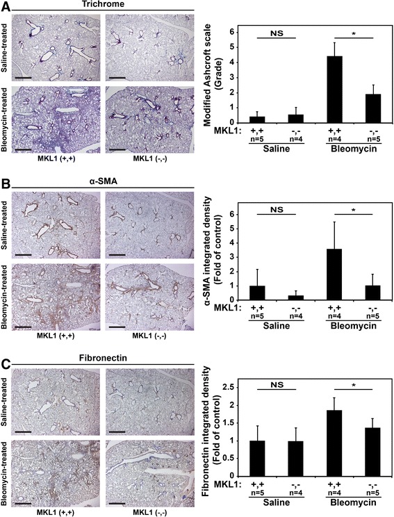
Figure 2.

MKL1 (−,-) mouse lungs following experimentally-induced fibrosis have decreased collagen, α-SMA and fibronectin accumulation compared to MKL1 (+,+) controls. A. Trichrome staining of MKL1 (+,+) and MKL1 (−,-) mouse lungs following bleomycin or saline treatment denoting areas of collagen accumulation (blue) with the corresponding modified Ashcroft score for each condition. B. Immunohistochemical staining for α-SMA (brown) and C. fibronectin (brown) including their respective quantitation. One-way ANOVA (p < 0.05) was used for statistical significance.
