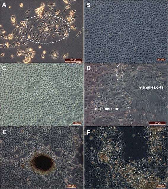
Figure 1.

Morphology of epithelial cells in follicular fluid. (A) Epithelial cells (inside dotted line) were found from one sample of follicular fluid after 3 days of culturing. (B) Epithelial cells had cobblestone-like morphology and grew rapidly after 7 days of culturing. (C) Epithelial cells were found from another sample of follicular fluid after 5 days of culturing. (D) To the left of the dotted line were epithelial cells and to the right were granulosa cells. (E) Human embryonic stem cell-like colonies were growing in the epithelial cell culture. (F) Contrasting with epithelial cells, granulosa cells appeared irregularly polygonal, and the pseudopodia were long and tightly adherent. Scale bars: 200 μm (A, D) and 100 μm (B, C, E, F).
