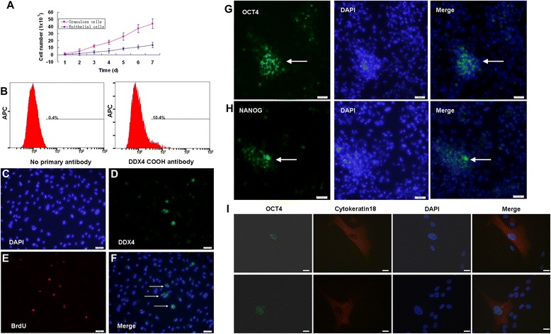
Figure 2.

Stem cell characteristics of follicular fluid-derived epithelial cells. (A) Typical cell growth curve of epithelial cells after seeding 2.5 × 10⁴ cells in each well of 24-well culture plates, compared with granulosa cells. (B) Cell-surface expression of DDX4 in epithelial cells was detected by fluorescence-activated cell sorting analysis after 14 days of propagation. (C), (D), (E), (F) Assessment of epithelial cells proliferation by dual detection of DDX4 expression (green) and bromodeoxyuridine (BrdU) incorporation (red) in vitro cultures. (G), (H) Part of the epithelial cells was OCT4-positive or NANOG-positive. (I) Double staining of OCT4 and cytokeratin 18 in epithelial cells. Scale bars: 100 μm (C, D, E, F, G, H) and 20 μm (I). DAPI, 4′,6-diamidino-2-phenylindole.
