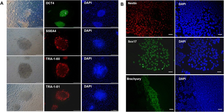
Figure 7.

Pluripotent characteristics of cell colonies derived from epithelial cell cultures using human amnion epithelial cells as a feeder layer. (A) Cell colonies showed positive immunofluorescence staining for pluripotency markers, namely OCT-4, SSEA4, Tra-1-60, and Tra-1-82. All cell colonies were from the same 2-month-old cell culture. (B) Cells were pluripotent, as demonstrated by their potential to differentiate in vitro into progeny representing the three germ lineages. Immunofluorescence staining showed differentiated cells expressing nestin (ectoderm), brachyury (mesoderm), and sox17 (endoderm). Scale bars: 200 μm (A) and 100 μm (B). DAPI, 4′,6-diamidino-2-phenylindole.
