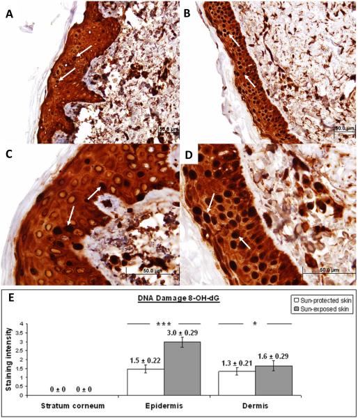
Figure 1. Immunohistochemical characterization and quantification of 8-OH-dG in UV-protected and UV-exposed human skin biopsy specimens.
A), C) UV-protected human skin sections stained with antibodies against 8-OH-dG. Strong nuclear staining is observed in approximately 15% of epidermal cells (arrows).
B), D) UV-exposed human skin sections stained with antibodies against 8-OH-dG. Strong nuclear staining is observed in approximately 90% of epidermal cells (arrows).
Moderate staining of intracelular material is observed equally in the epidermis of UV-protected as well as UV-exposed skin specimens.
Original magnification A, B × 40 and C, D × 100.
E) Analysis of staining intensity scores for 8-OH-dG. No difference was observed in 8-OH-dG staining of the stratum corneum of UV-exposed and UV-protected skin (0 ± 0 versus 0 ± 0). 8-OH-dG staining was significantly increased in the epidermis of UV-exposed versus UV-protected skin (3.0 ± 0.29 versus 1.5 ± 0.22; p = <0.01). 8-OH-dG staining was significantly increased in the dermis of UV-exposed versus UV-protected skin (1.6 ± 0.29 versus 1.3 ± 0.21; p = <0.05). *p < 0.05, ***p < 0.01. Data are shown as mean ± SEM; n=5.

