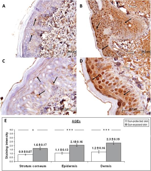
Figure 4. Immunohistochemical characterization and quantification of AGEs in UV-protected and UV-exposed human skin biopsy specimens.
A), C) UV-protected human skin sections stained with antibodies against AGEs. Weak staining is observed in epidermis (brown color staining, solid black arrow) and dermis (brown color staining, white arrow). Nuclear staining is demonstrated in approximately 10% epidermal cells.
B), D) UV-exposed human skin sections stained with antibodies against AGEs. Moderate staining is observed in the epidermis (brown color, black arrows) and dermis (brown color, long white arrow). Strong nuclear staining is noted in approximately 70% of epidermal cells (brown color, short white arrows).
The increased staining intensity in UV-exposed skin samples is determined to be 191% for stratum corneum, 196% for epidermis and 193% for dermis as compared to UV-protected skin samples. Original magnification A, B × 40 and C, D × 100.
E) Analysis of staining intensity scores for AGEs. AGEs staining of the stratum corneum was significantly increased in UV-exposed versus UV-protected skin (1.6 ± 0.17 versus 0.9 ± 0.07; p = <0.05). AGEs staining was significantly increased in the epidermis of UV-exposed versus UV-protected skin (2.1 ± 0.16 versus 1.1 ± 0.13; p = <0.01). HNE staining was significantly increased in the dermis of UV-exposed and UV-protected skin (2.3 ± 0.19 versus 1.2 ± 0.16; p = <0.01). * p < 0.05, ***p < 0.01. Student t test. Data are shown as mean ± SEM; n=5.

