. 2015 Apr 14;4:e06034. doi: 10.7554/eLife.06034

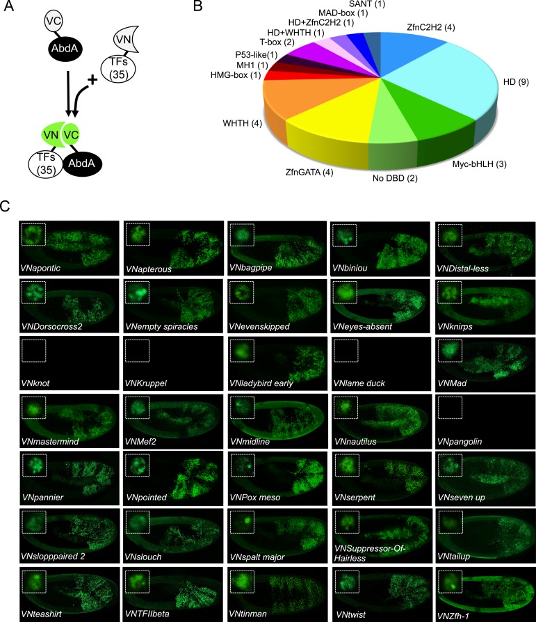
Figure 2. BiFC validates the AbdA-interaction status of competitive TFs.

(A) Principle of the BiFC screen between AbdA and the 35 selected TFs. (B) Repartition of the 35 selected TFs with regard to their DNA-binding domain. (C) Illustrative pictures of BiFC signals obtained between VC-AbdA and the indicated VN-TF in the epidermis of stage 10–12 of live embryos. Fusion constructs are expressed with the abdA-Gal4 driver. Note that typical nuclear interaction profiles are observed between AbdA and different TFs (white-dotted boxes). See also ‘Materials and methods’, Supplementary files 2, 3 and Figure 2—figure supplements 1, 2.

DOI: http://dx.doi.org/10.7554/eLife.06034.006

Figure 2.

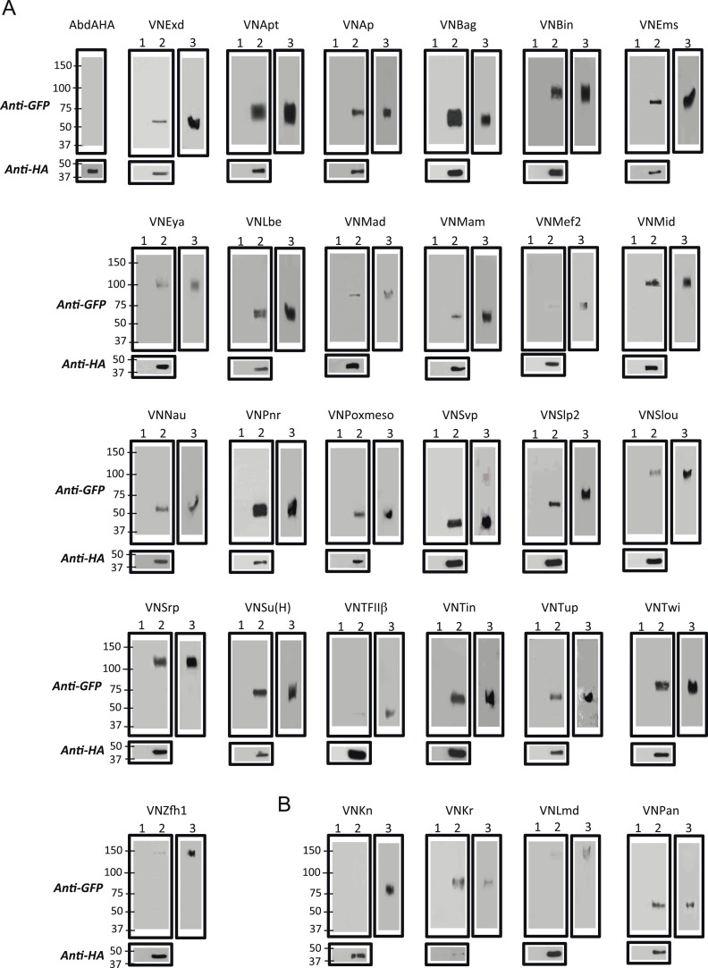
Figure 2—figure supplement 1. Co-immunoprecipitation (co-ip) between AbdA and TFs selected from the set used for BiFC in the Drosophila embryo.

Figure 2—figure supplement 1.

(A) Positive co-ip experiments. (B) Negative co-ip experiments. Co-ips were performed with an anti-HA antibody recognising the AbdA-HA variant. Presence of the TF was revealed with a polyclonal anti-GFP antibody recognising the VN fragment fused to each TF. The ip was performed with the fusion TF expressed alone (lane 1) or together with AbdA (lane 2). Lane 3 corresponds to the input of cells expressing both proteins (20% of the total lysate) before the ip. Gels on the bottom are western blots validating the efficiency of the ip with the anti-HA antibody. First gel corresponds to a control validating that the anti-GFP does not recognize AbdA-HA.
Figure 2—figure supplement 2. BiFC between mesodermal TFs and AbdA in the mesoderm.

Figure 2—figure supplement 2.

Fusion proteins were expressed with the 24B-Gal4 driver recombined to UAS-mCherry (red), which allows following the expression profile in the mesoderm. BiFC (green) was observed in stages 12–14 of live embryos. Note that weak BiFC signals could be observed with VN-Kn while VN-Lmd remained negative with VC-AbdA.