. 2015 Apr 14;4:e06034. doi: 10.7554/eLife.06034

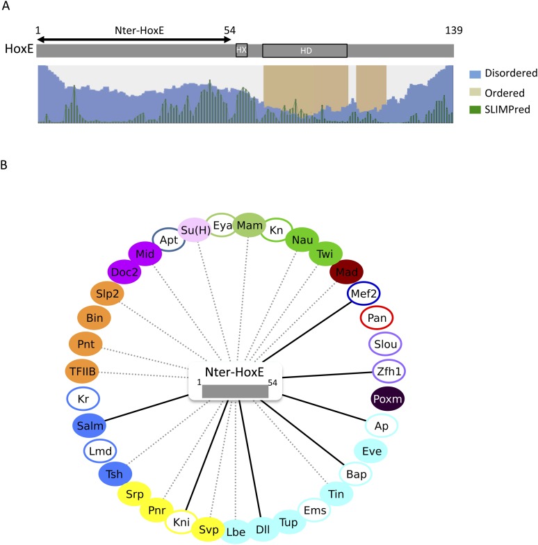
Figure 9. The intrinsically disordered region of HoxE establishes a number of interactions with Drosophila TFs.

(A) Scheme of full length Nematostella HoxE with its predicted SLiMs (green bars), and disordered (blue waves) or ordered (brown blocks) regions. Adapted from iupred (http://iupred.enzim.hu/). The N-terminal disordered region used for BiFC is indicated (Nter-HoxE). (B) Interactome between Nter-HoxE and the 35 Drosophila TFs. Colour code and representation are as in Figure 3. See also Figure 9—figure supplement 1.

DOI: http://dx.doi.org/10.7554/eLife.06034.036

Figure 9.

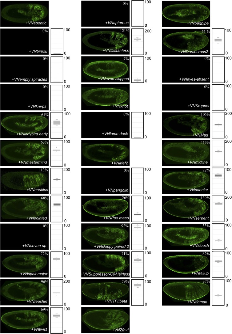
Figure 9—figure supplement 1. BiFC between Drosophila TFs and the N-terminal (1-54) fragment of the Nematostella HoxE protein.

Figure 9—figure supplement 1.

BiFC was performed with the 35 TFs in the epidermis of stage 10–12 embryos, as indicated. Quantification was performed as in Figure 3—figure supplement 1. No statistical quantification is provided in the case of a new interaction (as seen for Apontic, Bagpipe, Knot, Mef2 and Zfh1). No picture was taken in absence of BiFC.