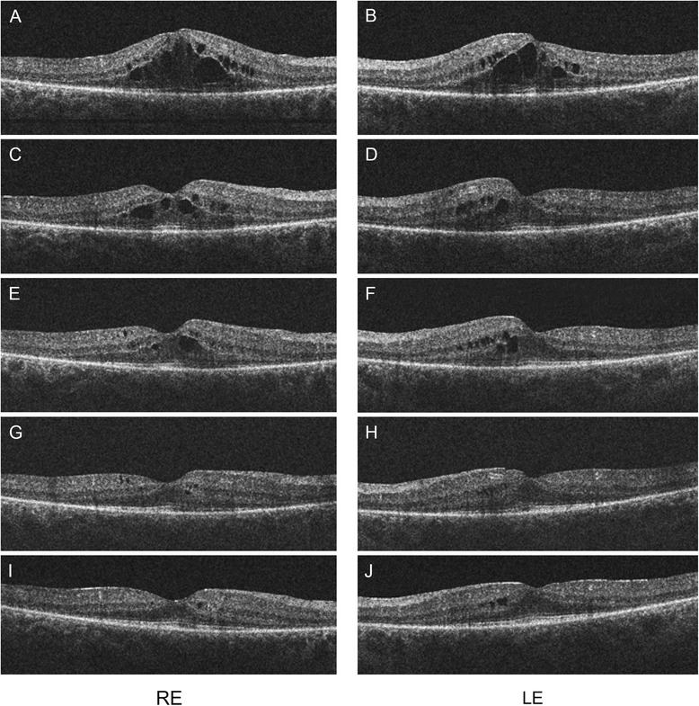
Figure 2.

OCT scans of the macula of both eyes after 4 series of TAC treatment (A, B). Large cystic spaces are present in the outer nuclear layer and small cystic spaces in the inner nuclear layer. Photoreceptor inner/outer segment (IS/OS) junction is discontinuous but relatively intact underneath the fovea and absent outside this area. Retinal thickness is lower outside the perifoveal area. Scans 5 weeks (C, D), 8 weeks (E, F), 12 weeks (G, H) and 17 weeks (I, J) after discontinuation of the chemotherapy. Retinal thickness in the perifoveal area gradually decreased along with the resolution of the intraretinal cystoid spaces.
