
Figure 2. Erk2 phosphorylates Drp1 Serine 616.
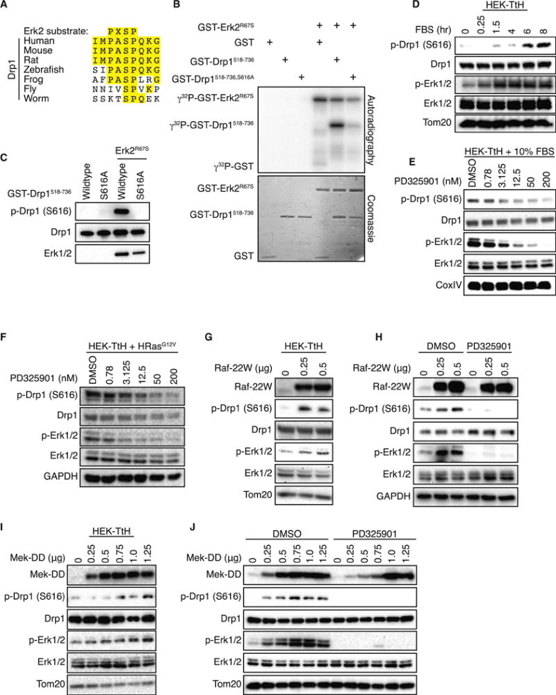
(A) Alignment of the consensus Erk2 target sequence with amino acids 612–620 of human Drp1 (isoform 1) and the corresponding sequence from the indicated species. (B–C) Recombinant, active GST-Erk2R67S was incubated with either GST alone, GST-Drp1518–736 or GST-Drp1518–736, S616A in the presence of γ32P-ATP (B) or ATP (C) and resolved by SDS-PAGE. Drp1 phosphorylation was detected by autoradiography (B) or immunoblot (A). (D–J) Phosphorylation of Drp1 (P616) and Erk1/2 (Y202/T204) were monitored by immunoblot in the following cells: (D) HEK-TtH cells grown in serum-free DMEM supplemented with 10μM Mek inhibitor PD325901 for 16 hrs, then supplemented with 10% FBS over an 8-hour time course; (E) HEK-TtH cells supplemented with 10% FBS and treated with 0.78–200nM of PD325901 for 8 hrs. (F) HEK-TtH cells stably expressing HRasG12V treated with 0.78–200nM of PD325901 for 8 hrs; (G) HEK-TtH cells transfected with increasing amounts of Raf-22W; (H) HeLa cells transfected with increasing amounts of Raf-22W in the presence of DMSO or PD325901; (I) HEK-TtH cells transfected with increasing amounts of MEK-DD; (J) HeLa cells transfected with increasing amounts of active MEK-DD in the presence of DMSO or PD325901; Tom20, CoxIV, GAPDH: Loading Controls.