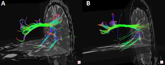
Fig 2. Example of DTI fiber tractography on one control (A) and one HDI (B).

Only three fiber bundles connecting the PCC/PCUN and MPFC, bilateral PHGs were detected in all the subjects. The color-coding of the obtained fibers is based on the standard RGB (Red, Green, Blue) code applied to the vector at every segment of each fiber. Red indicates the medio-lateral plane. Green indicates the dorsoventral orientation. Blue indicates the rostro-caudal direction. DTI = diffusion tensor imaging; HDI = heroin dependent individual; PCC/PCUN = posterior cingulated cortex/precuneus; MPFC = medial prefrontal cortex; PHG = parahippocampalgyrus.
