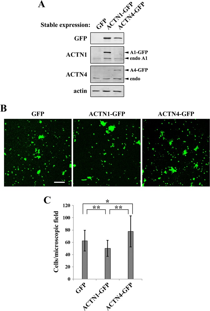
Fig 6. Invasion of DLD-1 cells expressing GFP, ACTN1-GFP, and ACTN4-GFP.
(A) Stable expression of GFP, ACTN1-GFP, and ACTN4-GFP in DLD-1 cells was examined by western blotting using the antibodies indicated to the left of each blot. (B) The invasion assay was performed using GFP-, ACTN1-GFP-, or ACTN4-GFP-expressing stable cell lines. Cells were seeded into Matrigel invasion chambers and allowed to invade into the Matrigel for 26 h. Then, the invaded cells were fixed and imaged by examining the GFP signals on the undersurface of the chamber using fluorescence confocal microscopy. Scale bar = 100 μm. (C) The GFP-positive cells shown in (B) were quantified. At least six microscopic fields obtained from duplicated chambers were used for quantification in each experiment. The results represent the mean ± SD of three independent experiments. *P < 0.05, **P < 0.01.

