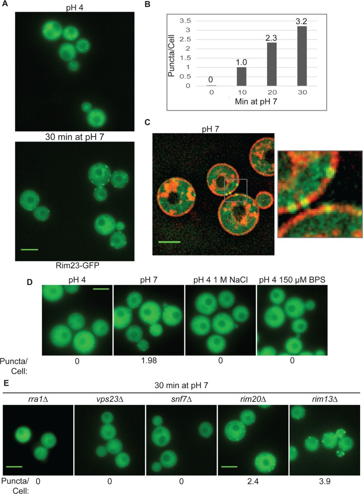
Fig 7. Rim23-GFP forms plasma membrane-associated puncta under neutral/alkaline pH conditions.
(A) Rim23-GFP was visualized at pH 4 SC McIlvaine’s buffer and after 30 min after shift to pH 7 SC McIlvaine’s buffer. (B) The number of Rim23-GFP puncta increase with time after shifting from pH 4 to pH 7. Quantification Rim23-GFP puncta/cell at pH 4 (0 min) and after 10, 20, 30 min incubation at pH 7. (C) Rim23-GFP puncta are closely associated with the plasma membrane. Cells were stained with FM-464 after 1 hr incubation at pH 7. (D) 1 M NaCl and 150 μM BPS does not induce Rim23-GFP puncta formation. Cells images after 30 min incubation in indicated culture media. (E) Rim23-GFP puncta formation was disrupted by rra1Δ, vps23Δ, and snf7Δ mutants. Cells were imaged after 30 min incubation at pH 7. The number of puncta/cell was quantified for 62–100 cells/strain. All strains in this figure were cultured in SC McIlvaine’s buffer media. All scale bars = 5 μm.

