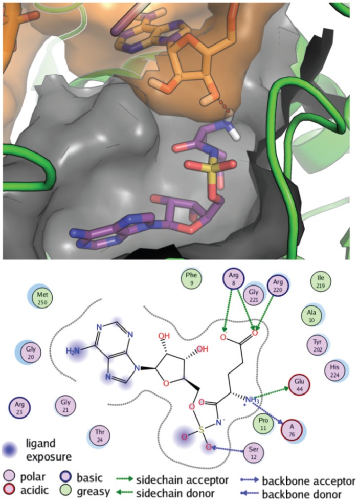
Fig 4. Molecular docking of Glu-AMS and E. coli tRNAGlu on E. coli GluRS.
Structural representation of the Glu-AMS and E. coli tRNAGlu in the E. coli GluRS binding site from the docking simulations. Top: Glu-AMS is in purple sticks, E. coli tRNAGlu A76 is in orange sticks and a transparent orange surface, E. coli GluRS is in green cartoon, and the binding site is depicted as a grey surface. The H-bond formed between Glu-AMS and A76 is shown as a dotted red line. Only the hydrogen atoms of the NH3 group involved in this H-bond are shown for clarity. Bottom: 2D representation of the Glu-AMS docked conformation bound to E. coli GluRS. The binding pocket is represented with a grey dotted line, polar and non-polar residues are represented as magenta and green circles, respectively, and polar interactions are shown as green and blue lines.

