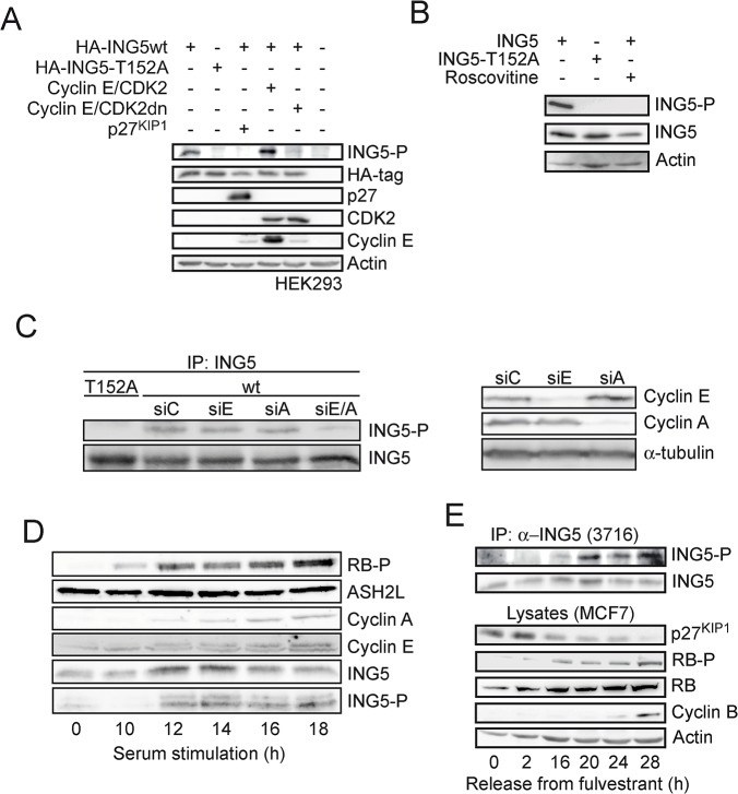
Fig 3. Phosphorylation of ING5 at T152 in cells.
(A) The indicated proteins were expressed transiently in HEK293 cells. T152 phosphorylation was assessed using mAb 3H5. The other proteins were detected as detailed in the Material and Methods section. (B) ING5 or ING5-T152A were expressed transiently in HEK293 cells and treated with 25 μM roscovitine 2 h prior to harvesting. ING5 and ING5-P were analyzed using mAb 7A11 and 3H5, respectively. (C) ING5 or ING5-T152A were expressed transiently in HEK293 cells. The cells were then reseeded and transfected with control siRNA (siC) or siRNA targeting cyclin E (siE), cyclin A (siA) or both cyclins (siE/A). ING5 proteins were immunoprecipitated using mAB 7A11 and T152-P and total amounts of ING5 were analyzed using mAB 3H5 and 7A11, respectively. Functionality of the siRNAS were analyzed in total cell lysates (right panel). (D) The cells of the glioblastoma cell line T98A were arrested in G0 with 0.1% FCS. Cell cycle re-entry was stimulated by reseeding and stimulating with 10% FCS. The expression of the indicated proteins was analyzed at the indicated time points after G0 release. T152 phosphorylation was measured using mAb 3H5.(E) MCF7 cells were released from a fulvestrant block (1 μM, 72 h) by adding a 10-fold excess of tamoxifen. The expression of the indicated proteins was analyzed at the indicated time points after addition of tamoxifen. Endogenous ING5 was immunoprecipitated (pAb 3716) and analyzed using mAB 3H5 and 7A11.

