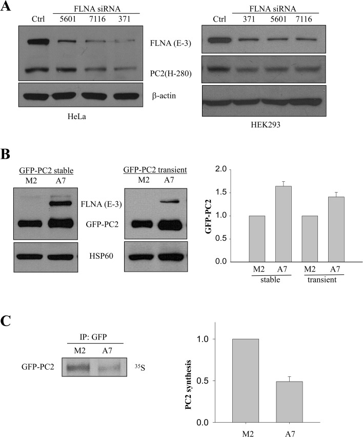
Fig 1. Effects of FLNA on PC2 protein expression and PC2 synthesis.
A, WB showing endogenous PC2 level in HeLa (Left panel) and HEK293 (Right panel) cells with FLNA KD by siRNA 5601, 7116 and 371, respectively. The numbers indicate the nucleotide positions in the FLNA mRNA open reading frame where the siRNA sequence starts. β-actin was used as a loading control. B, left panel, data from WB showing the expression of PC2 in M2 and A7 cells stably expressing GFP-PC2. GFP (B-2) antibody was used to detect GFP-PC2. Middle panel, PC2 expression in M2 and A7 cells transiently expressing GFP-PC2. Right panel, comparison between averaged GFP-PC2 levels normalized by HSP60 in M2 and A7 cells under stable and transient expression conditions. C, left panel, representative data from M2 and A7 cells showing GFP-PC2 synthesis assessed by 35S pulse labeling. Anti-GFP (EU4) was used to precipitate GFP-PC2. Right panel, comparison between averaged GFP-PC2 syntheses in M2 and A7 cells (N = 4; p = 0.004).

