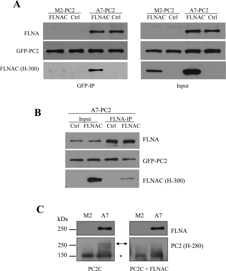
Fig 4. Effect of FLNAC on the FLNA-PC2 interaction.
All data are from three or more independent experiments. A, representative data from co-IP showing interaction of GFP-PC2 with FLNA and FLNAC in M2 and A7 cells. B, representative data showing the effect of blocking peptide FLNAC on the FLNA-PC2 interaction in A7 cells. C, Far WB showing the competition of FLNAC with PC2C for binding to FLNA. Lysates of M2 and A7 cells (stably expressing GFP-PC2) were separated by SDS-PAGE and transferred to nitrocellulose membrane. Proteins were denatured, renatured and then incubated with purified GST-PC2C and none (left panel) or His-FLNAC (right panel). Bound protein was detected by PC2 (H-280) antibody. The arrow (←) indicates PC2C signal detected at the site of FLNA. The star (*) indicates stably expressed GFP-PC2 signal, as a control.

