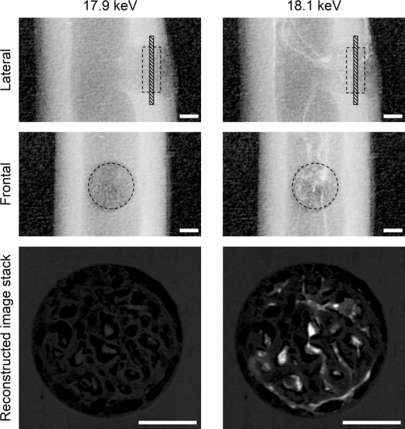
Figure 1.

Lateral and frontal radiographs and stacks of reconstructed images of the drill-hole defect from a WB10 rat, obtained from synchrotron radiation at 17.9 keV (left) and 18.1 keV (right). The reconstructed image stacks comprise twenty 2.74-μm-thick slices perpendicular to the drill-hole direction (hatched regions in the lateral radiographs). The enhanced contrast between blood vessels and bone on the 18.1-keV images is caused by the sharp absorption jump of ZrCA at just above its K-edge. The intercortical dashed circles and squares indicate the drill-hole defect region for analysis. Bar: 300 μm.
